Wednesday, March 29, 2017

Pics! Videos Coming Soon as Well!

Because a picture is worth a thousand words.

Results from the neuroanatomical imaging experiment. The green denotes the Rdl GABAa receptors, and the other colors are from the neurobiotin dye injections. As you can see, the dye makes different parts of the brain show up in various shades of pink and orange, and the receptors glow green from the immunohistochemistry. Based on overlaps/no overlaps of these colors, you can tell what parts of the brain, and what neurons in the brain, have/don't have the receptors. These are images from the confocal microscope.


I don't know how to film videos through a microscope, or do a surgery one-handed while filming with the other, but I can still take pics of what I see. These are images of moments during my practice surgeries. As you can see from these images, we trap the bees in a different harness for surgeries, and we open the head to do injections, before closing them again. I was lucky this particular bee was a practice one - it died on me (you will be remembered, random bee).


In the neuroanatomical experiment, during the immunohistochemical portion, you have to take thin cross-sections of the brain, after you fix them in agarose. This is a pic of me next to the vibratome used to take the sections. I have a video of me actually collecting sections with this device - will post videos soon.


The procedure for RNA Isolation by Trizol/Chloroform Extraction and Isopropanol Precipitation.


Preparing the centrifuge during the RNA Isolation procedure.


Sometimes, during the RNA Isolation procedure, you have to briefly mix the samples with a smaller centrifuge.


A pic of my samples in the middle of the RNA Isolation procedure, next to a bottle of the Trizol reagent I use during the process. Trizol is quite dangerous - it causes severe skin burns, and it's also toxic if you inhale it, so I always take care to wear gloves and a lab coat, and to work inside the fume hood.


Before we did RTq-PCR, we had to measure the concentration of RNA in our samples using a spectrophotometer. This spectrophotometer is not like the ones from school, as all it needs is one drop. We used this technology to check for contamination in our samples.


Procedure for preparing samples for RTq-PCR:


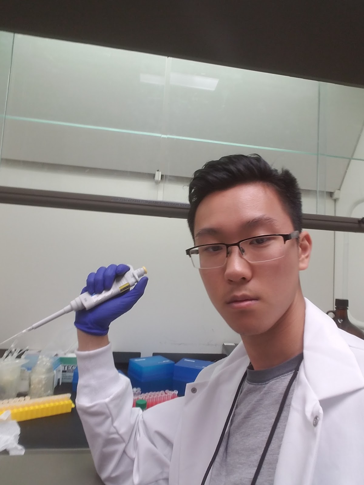
Micropipetting diligently (you can almost see the exhaustion in my face). It hurts your neck if you micropipette for a while - I always take full advantage of my lunch breaks to stretch and move around.


Listening to music while waiting for the centrifuge to do its job:


Week 8: Truth

It's finally coming together.

So I finally found out answers to a TON of my questions - the most important one being, how is all this molecular bio stuff related to the neuroanatomy side I was working on about half a month ago?

It all starts with the Rdl subunit. The inhibitory GABAa receptor in the honey bee has subunits, and they work together to get the receptor to work. Ligand-gated ion channels are found in all these subunits, and all the subunits should be important to signaling. The Rdl subunit is one such subunit.

When the lab did the genetic and molecular biology side of this experiment (which is still ongoing, I did another round of RNA Isolation this week), the lab injected RNA into the bees to affect expression of the proteins needed to make the Rdl subunit. There were three treatments - dsiRNA (Dicer-substrate short interfering RNA), scrambled RNA, and non-injected. The dsiRNA specifically interferes with the sequence in question, called AmelRdl, that codes for the receptor proteins, preventing its transcription. Scrambled RNA is just nonsense RNA that shouldn't have any effect on expression. And the non-injected group just didn't get any injections at all, so it's a control group.

So the injections happened, and then, the actual experiment commenced (Note: These injections are completely different from the injections I did during the neuroanatomical experiment - that time, I was injecting neurobiotin dye for imaging). All three groups were subject to PER (Proboscis Extension Reflex) conditioning experiments, and then the bees were analyzed, via RNA Isolation and RTq-PCR, to check for levels of the AmelRdl RNA sequence in question after conditioning. This sequence codes for the receptor proteins in question, so correlating levels of this RNA sequence to observed behavior can give us conclusions about the role of the Rdl proteins, and thus, the subunit.

This brings me to the answer to another question I had: Why RNA?

If we do regular PCR on the bees, we get amplification of the bee's entire genome, regardless of whether parts of the genome are being expressed or not. The experiment cares about tracking expression of the right sequence, which would mean we only care about the parts of the genome being expressed. That's why we collect RNA - RNA is present when those corresponding parts of the genome are being expressed. And in the collected RNA, there should be the AmelRdl RNA sequence.

So now, the connection with the neuroanatomical side. I looked at our poster again today, and looking at the conclusions at the bottom of the poster, I noticed that it said the AmelRdl receptors aren't found in the olfactory receptor neurons.

The conclusion I said from the neuroanatomical experiment about the GABAa receptors and the olfactory receptor neurons was basically the TL;DR version of the more specific conclusion - the Rdl subunit isn't found in those neurons. So the entire experiment is not only based around a specific receptor, but is based around a specific subunit in it.

So where am I now in this experiment? Like I stated above, this week, I did another round of RNA Isolation, this time, for the GABAa receptor experiment. Mary's experiment concluded last week (refer back to last week's post if you don't know who I'm talking about), so now I'm doing this procedure for my experiment. Giulia (the other undergrad in my project) provides the samples, and I analyze them. Next week, Dr. Sinakevitch said I'd be doing the RTq-PCR process alone for my samples. This should be pretty fun - will get tons of pics. She'll probably have me practice micropipetting again before I start though - micropipetting for RTq-PCR is extremely precise - I have to micropipette a drop of about 2 microliters onto the side of a sample well about 2 millimeters in diameter. Not even on the top. Bummer - another morning of dull practice.

And...I just remembered about the images over the last few weeks. I will get them up right now.


Thursday, March 23, 2017

Weeks 6 and 7: Intro to Molecular Biology

Weeks 6 and 7 have been great! I made a ton of progress in the lab, and I got to do a TON of practical procedures. I am excited about weeks 8 and 9 now.

The highlight of these weeks was learning a new procedure - RNA Isolation by Trizol Extraction and Isopropanol Precipitation. A mouthful, but for anyone who wants the TL;DR version, it means extracting genetic material from samples of honey bee brains. DNA and RNA are both extracted from the procedure, and the lab then specifically isolates the RNA (Why do we only care about the RNA? I still am not entirely sure).

Why do we care about this procedure? On the deliverable poster we submitted to the 2017 Arizona Imaging and Microanalysis Society's Microscopy Conference, there was a section mentioning levels of RNA. The lab was conditioning bees with the PER (Proboscis Extension Reflex), and isolating the RNA sequence used to make the GABAa receptor from these conditioned bees, in order to make correlations with the observed behavior and the amount of the RNA sequence isolated. So the lab was trying to find out if high or low levels of the GABAa receptor RNA code could be correlated to the behavior observed from conditioning trials.

This avenue of our research is still ongoing, and I probably will be doing this for the GABAa receptor over the next few weeks. These last two weeks, however, I did this procedure for a different receptor, the tyramine receptor. It's the same procedure, just a different receptor in question. It wasn't my project - I was assisting the graduate student, Mary Peterson, in her research for her Master's degree thesis. Dr. Sinakevitch had me do this to give me the experience I need to have this procedure down. After having me practice micropipetting for a morning, I was ready to go.

Molecular biology requires much precision to get truly accurate results - even a microliter makes all the difference. I had to be really careful the entire time while I was doing the procedure. The micropipetting practice definitely helped, but I had to be careful of a ton of other things too, notably, contamination of the samples and timing for steps such as centrifuging. I thought for sure I was going to mess up something along the way. But I can surprise even myself - the start of Week 7, I did the entire first part of the procedure alone for a dozen samples, no errors.

I was proud of myself - the only real practice I had had was doing some steps of the procedure alongside Mary and Dr. Sinakevitch in Week 6. But the real fun started after I did the procedure alone.

Dr. Sinakevitch showed me what happens next in the second half of Week 7. It turns out that what I had done so far was the first big part of a larger process. I had extracted both DNA and RNA, but I had to separate the RNA. She showed me how to use the kits for separating the RNA, and soon, we were ready for the final part. It was a good thing I did the first part really well - it messes up everything afterwards if you mess up there.

With our small amounts of RNA, we can't really make any conclusions. We needed to amplify our samples. That's what the last part, RTq-PCR, is for. Reverse-transcription Quantitative Polymerase Chain Reaction. This takes the RNA, builds complementary DNA strands, and then amplifies the DNA built, which will show the amount of RNA we got. I'm sure there are more important details associated with this procedure, but I haven't done this last part yet - I only watched Dr. Sinakevitch do it. She said I'd be doing this soon, though, so once I do it, I can get back to all of you on this.

My thoughts on these last few weeks? Amazing. It's getting difficult these days to keep track of everything I know - I welcome the challenge though. This is more biology then I've ever been exposed to. By the time this project is over, I'll be ready to continue doing more of this in a college environment. Also, after doing the RTq-PCR, Dr. Sinakevitch told me the samples I did were excellent, so now, I am very confident going into weeks 8 and 9. I'm just hoping I get to do the RTq-PCR procedure in the next two weeks - it looks really exciting.

Also, I just realized I should post some pics of what's been going on. I'll do that this weekend - the visuals of the last few weeks are too good not to share.

Friday, March 10, 2017

Weeks 4 and 5 in the Lab

In Week 4 in the Smith Lab, I continued learning more about the neuroanatomical side of the experiment with Dr. Irina Sinakevitch - I needed to get familiar with the procedure, and I needed to understand our results.

In the week before (Week 3), Dr. Sinakevitch and I had taken 5 bees and injected the fluorescent dye into specific areas of their brains, notably, the antennal lobes and the projection neurons, so that these areas would show up in confocal imaging. During Week 4, I continued my practice of this injection procedure, specifically, the surgery needed to open the bee's head for injections. For my Week 4 practice, I took 2 new bees, one on Monday and one on Tuesday. Unfortunately, when I practiced on those two bees, I failed the surgery both times. I accidentally pushed my razor too far into the head for one of the bees, and I failed to make the proper incisions in the other bee's head, meaning I couldn't close that bee's head again. The locations of the incisions are hard to see, and the bee brain is a very delicate structure - I needed more practice and experience before I could get this procedure down. My first-time success in Week 3 was beginner's luck, I guess. Good thing too - Dr. Sinakevitch and I were able to use that lucky bee brain for imaging, alongside the four other good ones Dr. Sinakevitch had performed surgery on then, so we were still able to use all 5 bees from Week 3 to get proper results. During Week 4, while I practiced the injection procedures, Dr. Sinakevitch did the immunohistochemistry to label the GABAa receptors in our Week 3 samples, and we were set to image.

When we imaged the brains from Week 3 with confocal microscopy that week, we got results. Dr. Sinakevitch had used immunohistochemistry to label the GABAa receptors with antibodies after we had done the injections. Due to both the fluorescent antibodies and the fluorescent dye, we were able to light up the parts of the brain we cared about with one color, and the locations of the GABAa receptors within those parts showed up as another color. With this procedure, we were able to deduce that the GABAa receptors were not located in the olfactory receptor neurons, which are in the antennal lobes. This result was the main result we communicated in the deliverable poster we made for the 2017 Arizona Imaging and Microanalysis Society's Microscopy Conference, which was that Friday. I spent the last day of Week 4 assisting Dr. Sinakevitch in editing the poster, and I even got to show her some tricks on Microsoft Word while we were writing parts of our poster. Unfortunately, I couldn't go on Friday - I was in UT Austin at the time for a college orientation. My editing did help a ton for the poster though, as my mentor is not completely fluent in the English language and needs someone to help with her grammar. I was also very proud that my only surgical success amounted to something, and relieved that I could understand what was going on. And I was super happy to be second author on that poster - that poster is hanging now on the walls of ASU's ISTB (Interdisciplinary Science and Technology Building) 1. 

Week 5 was when I finally got to see the immunohistochemical side of our neuroanatomical experiments. Of course, I also kept practicing the surgical injection procedure that comes before. In Week 4, though we had gotten results from the olfactory receptor neurons, we had not gotten results from the projection neurons - we were not able to use the images. Monday of Week 5, we took five bees, this time, to image them for results from the projection neurons. Dr. Sinakevitch opened the head and injected for three bees, and left me two to practice with. Very generous. 

"I couldn't fail this time," I thought as I got the materials ready. My thoughts didn't matter - pretty soon, I lost another bee, and I was left with just one more. 

I proceeded as carefully as I could with that bee, and this time, I succeeded in injecting and closing the head again. The bee had survived the procedure. How relieved I was! I gave Dr. Sinakevitch that bee so she would have another brain to use alongside her three. The next day, however, I was informed that my bee died shortly after I gave it to her. We were down to three bees. 

Depressing, but this is where it begins to get better. Dr. Sinakevitch had gotten her three brains out, and she decided it was time I learned the immunohistochemical procedure. I learned how to prepare the agarose gel and how to block our brains in the agarose, before learning how to use the vibratome to take sections of our brains. I got to take sections for two of the three brains, and I got it perfectly. It came easy - much better than the surgery. Next. Dr. Sinakevitch showed me the procedure of washing the sections, to prepare them for the primary antibodies. This means applying the PBS (phosphate buffer solution), with the detergent in it, to the sections at least 6 times. I did this part too. Time management was crucial for this step, because taking out and putting the solution had to be timed, but luckily, I've always liked being punctual. After the first application of the solution for one hour, the next applications had to be for 10 minutes each. There are other ways to do this timing, but Dr. Sinakevitch said this timing was okay. Dr. Sinakevitch came in after that and applied the primary antibodies, and our sections were ready. There are steps after this, which I was supposed to see on Wednesday, but I fell sick on Wednesday with a flu, and I couldn't come in. But it's okay - I still have videos of me doing the washing and the vibratome sections. I also got pics of our poster, and the confocal images I saw. 

My thoughts? Much of this is still quite complicated. I am still adapting to the lab. Despite this, however, I am learning so much, maybe more than I ever have before, and I am already making my impact. And I'm doing okay - I can do the immunohistochemical steps, and I think now, I can succeed when I do the surgery again. I almost succeeded on that last bee I performed surgery on - next time, I know it'll be better. I am confident I can go forth in neuroscience and neurobiology after this, carrying new abilities and experiences from this lab that will definitely come in handy.